Anatomy Of Upper Leg Muscles And Tendons / Upper Leg Pain Kinesiology Sports Tape / The muscles that form the quadriceps femoris unite proximal to the knee and attach to the patella via the quadriceps tendon.
Anatomy Of Upper Leg Muscles And Tendons / Upper Leg Pain Kinesiology Sports Tape / The muscles that form the quadriceps femoris unite proximal to the knee and attach to the patella via the quadriceps tendon.. The thigh is the area between the hip and the knee joint. The triceps tendon is wider than most of the other tendons in the upper extremity. Rectus femoris these four muscles come together to form a single tendon, which inserts into the patella, or kneecap. Anatomy of the hand and wrist: It runs from your inner thigh to the quad tendon.
This is the group of muscles that you often see body builders flexing, which protrude just above the knee and take up most of the upper leg. Possibly the most important tendon in terms of mobility is the achilles tendon. Its muscle belly is on the back aspect of the upper arm. A tendon connects the muscle to the bone. The wrist links the hand to the forearm.
The Leg Ankle And Foot Amboss from media-us.amboss.com One of the most important tendons in terms of mobility of the leg is the achilles tendon. Rectus femoris muscle, one of the. The thigh is the area between the hip and the knee joint. It is part of the lower limb. The triceps tendon is wider than most of the other tendons in the upper extremity. Tendons vary in size and are somewhat elastic and attach bones to muscles. On the anterior side, the most prominent of the muscles are the sartorius muscle and the four muscles that make up quadriceps muscle group (the quads.) This important tendon in the back of the calf and ankle stores the elastic energy needed for running, jumping, and other physical activity.
The wrist is a complex system of many small bones (known as the carpal bones) and ligaments.
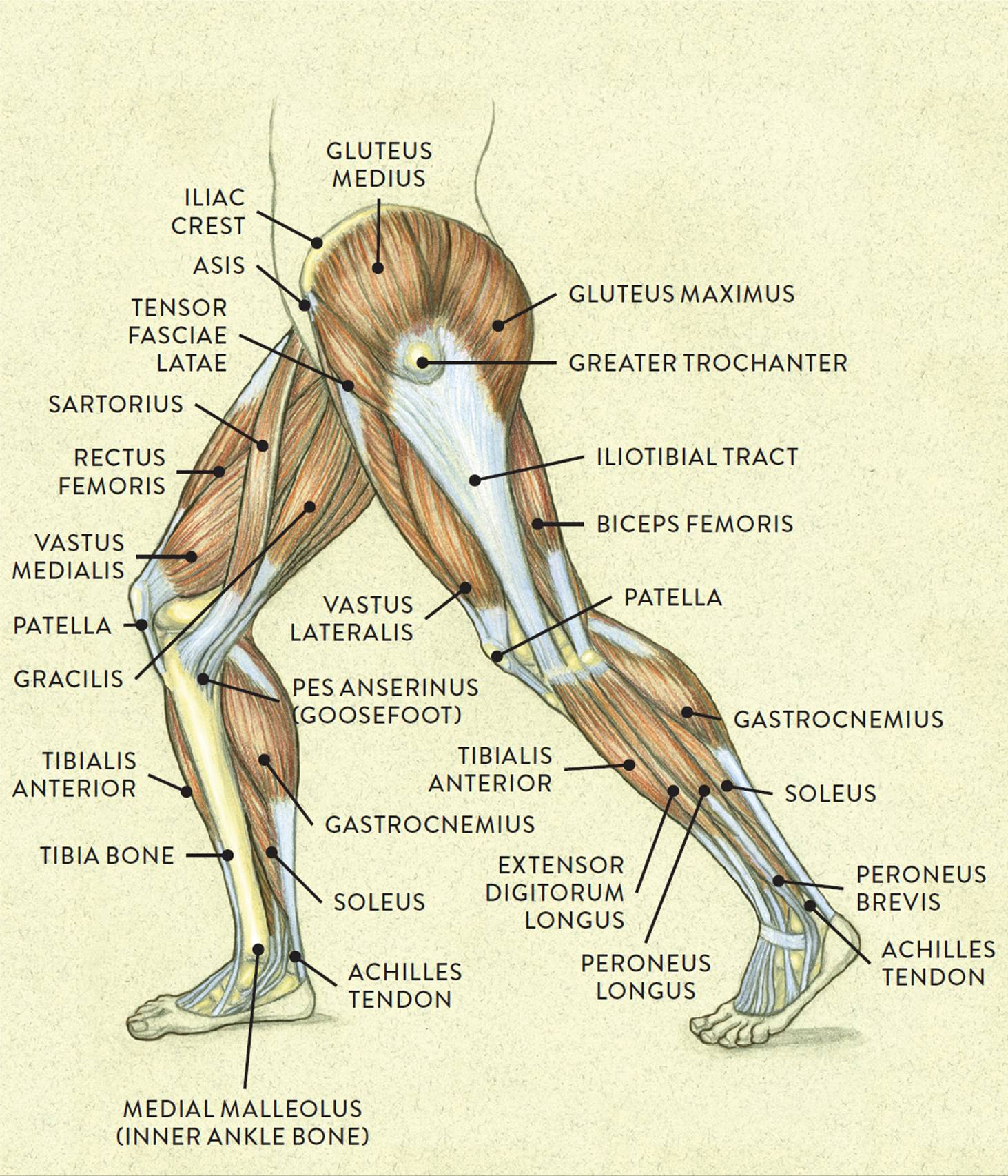
The biceps femoris hamstring muscle is the most frequently injured, as it suffers the largest stretch during sprinting, followed by the semitendinosus muscle. In turn, the patella is attached to the tibia by the patella ligament. Über 7 millionen englischsprachige bücher. Biceps femoris it allows your knee to flex and rotate and your hip to extend. They form the main bulk of the thigh, and collectively are one of the most powerful muscles in the body. Tendons attach muscle to bone. They consist of the rectus femoris, vastus intermedius, vastus lateralis and the vastus medialis. On the anterior side, the most prominent of the muscles are the sartorius muscle and the four muscles that make up quadriceps muscle group (the quads.) Tendons attach the muscles to each other. This anatomy chart is a great example of beauty and function in one, as it is pleasing to look… There are 3 muscle bellies that join to make this tendon. Schau dir angebote von muscle anatomy auf ebay an. To bend the elbow and to turn the palm of the hand towards the sky.
They consist of the rectus femoris, vastus intermedius, vastus lateralis and the vastus medialis. The single bone in the thigh region is called the femur. It forms a tendon near the elbow. The biceps femoris hamstring muscle is the most frequently injured, as it suffers the largest stretch during sprinting, followed by the semitendinosus muscle. Related posts of muscles and tendons of the leg muscle anatomy for gym.
Muscles Of The Leg And Foot Classic Human Anatomy In Motion The Artist S Guide To The Dynamics Of Figure Drawing from doctorlib.info The carpal bones are arranged in 2 interrelated rows. Über 7 millionen englischsprachige bücher. This important tendon in the back of the calf and ankle stores the elastic energy needed for running, jumping, and other physical activity. Tendons vary in size and are somewhat elastic and attach bones to muscles. Ligaments and tendons are fibrous bands of connective tissue that attach to bone. Soft tissues called tendons connect these muscles to the bones of the pelvis, knee, and lower leg. A tendon connects the muscle to the bone. One row connects with the ends of the bones in the forearm—the radius and ulna.
The wrist links the hand to the forearm.
The single bone in the thigh region is called the femur. The gastrocnemius muscle supersedes its function. They consist of the rectus femoris, vastus intermedius, vastus lateralis and the vastus medialis. The triceps tendon is wider than most of the other tendons in the upper extremity. This is the group of muscles that you often see body builders flexing, which protrude just above the knee and take up most of the upper leg. The largest tendon in the knee is the patellar tendon. The wrist is a complex system of many small bones (known as the carpal bones) and ligaments. It forms a tendon near the elbow. They are attached to the femur (thighbone), tibia (shinbone), and fibula (calf. It is part of the lower limb. One of the most important tendons in terms of mobility of the leg is the achilles tendon. This anatomy chart is a great example of beauty and function in one, as it is pleasing to look… One row connects with the ends of the bones in the forearm—the radius and ulna.
Tendons attach the muscles to each other. Rectus femoris these four muscles come together to form a single tendon, which inserts into the patella, or kneecap. The single bone in the thigh region is called the femur. The gastrocnemius muscle supersedes its function. The quadriceps muscles provide strength and power with knee extension (straightening).
Thigh Muscle Diagram Leg Muscles Diagram Muscle Diagram Leg Muscles from i.pinimg.com The muscles that affect the knee's movement run along the thigh and calf. The tendons have 2 functions: Über 7 millionen englischsprachige bücher. The biceps femoris hamstring muscle is the most frequently injured, as it suffers the largest stretch during sprinting, followed by the semitendinosus muscle. When the muscle contracts, the tendons are pulled, and the bone is moved. Related posts of muscles and tendons of the leg muscle anatomy for gym. Other muscles of the anterior (front) thigh include the pectineus, sartorius,. Ligaments and tendons are fibrous bands of connective tissue that attach to bone.
This important tendon in the back of the calf and ankle connects.
Rectus femoris muscle, one of the. The quadriceps muscles provide strength and power with knee extension (straightening). The carpal bones are arranged in 2 interrelated rows. Iliopsoas muscle, a hip flexor muscle that attaches to the upper thigh bone. Included are several layered views of the back muscles, the dorsal muscles, subclavius muscles, rhomboideus major and minor muscles, deltoid muscles and many more. Your upper leg includes seven major muscles. The muscles that affect the knee's movement run along the thigh and calf. The wrist links the hand to the forearm. Attachment, nerve supply & action. Muscle anatomy for gym 12 photos of the muscle anatomy for gym muscle anatomy and fitness, muscle anatomy for fitness, muscle anatomy for gym, human muscles, muscle anatomy and fitness, muscle anatomy for fitness, muscle anatomy for gym There are 3 muscle bellies that join to make this tendon. They form the main bulk of the thigh, and collectively are one of the most powerful muscles in the body. It is part of the lower limb.
The gastrocnemius muscle supersedes its function upper leg muscles and tendons. The muscles that form the quadriceps femoris unite proximal to the knee and attach to the patella via the quadriceps tendon.


0 Comments